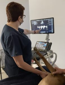
At IVC, we offer diagnostic ultrasounds with an outpatient, appointment-based schedule.
What is an ultrasound?

An ultrasound examination, also known as ultrasonography, is a non-invasive imaging method that we use to see internal organs by recording echoes or reflections of ultrasonic waves.
Ultrasound equipment directs a narrow beam of high frequency sound waves into the area of interest. The sound waves may be transmitted through, reflected, or absorbed by the tissues that they encounter. Bone and air will both stop ultrasound waves, so we mainly use ultrasound for body regions and organs that are not surrounded by bone or filled with air.
Ultrasonography has clear advantages over radiography (x-rays) when evaluating abdominal organ abnormalities, non-abdominal soft tissue problems, fluid build-up, heart disease, and many others. We also use ultrasound for non-diagnostic reasons, including safely guiding needles for sample collection.
Your veterinary may recommend an ultrasound in order to evaluate:
- Unexplained weight loss
- Persistent diarrhea or vomiting
- Abnormalities on abdominal palpation
- Pregnancy
- Fluid build-up in the abdomen or chest
- Abnormal blood work results
- Chronic infections
- Abnormal urinary habits
- Enlarged heart on chest x-rays
- Suspected heart failure
- Ligament or tendon tears
- Pre-surgical assessment
What will happen at my pet’s appointment?
Preparing for a successful ultrasound starts at home! In most cases, we recommend having your pet skip breakfast on the morning of the ultrasound. If we are performing an ultrasound of your pet’s abdomen, having them skip breakfast helps us to get the best possible images of the stomach and the area around it (since the stomach is empty during the exam). Your veterinarian may have prescribed an anti-anxiety medication for your pet to take the morning of their ultrasound. If so, it is okay to give this medication with one small meatball of food. We also recommend discouraging your pet from urinating (as much as that is possible) within the two hours before your appointment. If your pet’s bladder is full at the time of the ultrasound, it is easier to obtain accurate ultrasound images of the bladder wall.
Before your appointment, Dr. Mueller and her team will review your pet’s medical records. Your pet will also receive a full physical examination before their ultrasound. The ultrasound exam is painless, but your pet must be very relaxed in order to obtain diagnostic images. We will place an intravenous catheter in order to administer a mild sedative (if needed) in order to enhance relaxation.
We will clip the fur over the area on your pet that we plan to examine by ultrasound. This is because the sound waves that create the diagnostic images obtained by ultrasound do not travel through air. Hair traps air and disrupts the sound waves, which affects the clarity of ultrasound images. Clipping the area of interest and applying a thin layer of gel improves contact between the patient’s skin and the ultrasound probe, which enhances the quality of images.
Remember, ultrasound waves must travel into and out of the patient efficiently in order to produce the most diagnostic image possible! This means we can obtain the most possible information about your pet’s health condition.
Depending on the ultrasound findings, Dr. Mueller may recommend an additional blood test. She may also recommend obtaining samples of an abnormal internal organ using a small ultrasound-guided needle (for an aspirate) or biopsy instrument (for a biopsy). This requires a brief anesthesia, in order to make sure your pet is fully relaxed and stays as still as possible. The results of these tests are usually available within a few days.
An ultrasound appointment typically takes about an hour to an hour and a half, and we will send the results of your pet’s ultrasound to your primary care veterinarian after the appointment is complete.